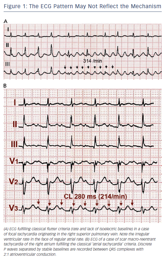
Ablation of this rhythm targets a critical portion of the circuit know as the cavotricuspid isthmus (CTI) it is to this area bounded by the tricuspid valve and the inferior vena cava (IVC) that a linear ablation lesion can be delivered with successful interruption and termination of the circuit. Animated illustration of the electrical circuit in the right atrium during typical atrial flutter. Conditions that lead to right-sided heart dilation and increased pressure can increase the incidence of this rhythm disturbance (right-sided heart failure, pulmonary hypertension, COPD, hypertension, obesity, tricuspid or pulmonary stenosis).įigure 1. It can occur in patients with no structural heart disease (lone atrial flutter), but is more likely in those with a history of prior cardiac surgery, congestive heart failure, or congenital heart disease. The heartbeat on exam is typically fast, but with medications, it can be slow with either a regular or irregular pulse.Ītrial flutter is the result of a rapid electrical circuit localized to the top right heart chamber, the right atrium. This electrical circuit has been extensively mapped using electrophysiology-guided catheter mapping techniques. The atria can contract anywhere from 250-400 beats per minute, with the AV node serving as the “traffic control” preventing 1 to 1 conduction to the ventricles. Treasure Island (FL): StatPearls Publishing 2022 Jan-.Atrial flutter, another type of supraventricular tachycardia, produces a heart rhythm with typically more atrial contractions than ventricular beats.

Now test your knowledge on this material by taking the atrial flutter ECG quiz.Īmerican Heart Association | Algorithms. This is a procedure that ablates (destroys or erodes) some of the tissue in the heart to prevent it from firing irregularly. In some cases, if the rhythm is persistent an ablation may need to be performed. If the patient becomes unstable where the blood pressure is dropping, mental status changes are occurring, chest pain, shortness of breath (signs indicating decreased cardiac output), a synchronized cardioversion may be necessary. Therefore, the patient may need some type of anticoagulant like “ Warfarin” etc. In addition, it’s important to prevent blood clot formation. To do this, medications can be used such as, calcium channel blockers (Diltiazem), beta blockers (propranolol) or Digoxin (if patient has heart failure) along with antiarrhythmics (Amiodarone). The treatment is similar as a-fib in that you want to control the rate. Heart valve problems like with the tricuspid or mitral valves, overactive thyroid, can present after heart surgery or a heart attack Treatment for Atrial Flutter

Therefore, it’s important the rate is managed, hence under control.


These waves have a saw-tooth appearance to them. P-waves are NOT present, but abnormal p-waves that are termed flutter waves.The atria are firing very rapidly with an atrial rate of around 300 bpm.The atrial rate is regular.No p-waves but flutter waves that look like a saw tooth.


 0 kommentar(er)
